The folding graph visualizations
A folding graph is a connected component of a protein graph. Here you can search for all visualizations of the different folding graph types of a protein chain. For each of the folding graph types (e.g., the alpha graph, which only considers alpha helices),
there are four different visualizations available:
- the ADJ notation: all SSEs of the protein graph are shown, order is from N-terminus (left) to C-terminus, and each edge represents a 3D contact between a pair of SSEs.
- the RED notation: only SSEs of the folding graph are shown, order is from N-terminus (left) to C-terminus, and each edge represents a 3D contact between a pair of SSEs.
- the SEQ notation: all SSEs of the protein graph are shown, order is from N-terminus (left) to C-terminus, and each edge represents the sequential order in the folding graph.
- the KEY notation: only SSEs of the folding graph are shown, order is spatial, and edges follow the SSEs in sequence order (N to C terminus). Note that it is not possible to define a spatial ordering for bifurcated graphs, so such graphs do not have a KEY notation.
Search Results
| FG# | Fold name | # SSEs | SSE string (N to C) | First vertex # in parent PG | Notation adj | Image available | Linnot overview page |
|---|---|---|---|---|---|---|---|
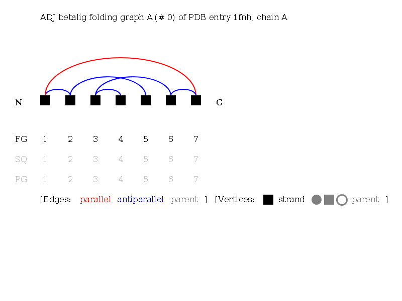
| 0 | A | 7 | EEEEEEE | 4 | (e,-1ae,3ae,1ae,-6pe,1ae,3ae,-3ae) | Fold A | Go to linnots |
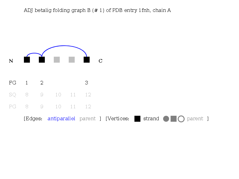
| 1 | B | 3 | EEE | 8 | [e,1ae,3ae] | Fold B | Go to linnots |
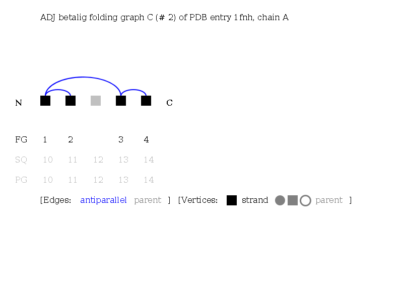
| 2 | C | 4 | EEEE | 11 | [e,-1ae,3ae,1ae] | Fold C | Go to linnots |
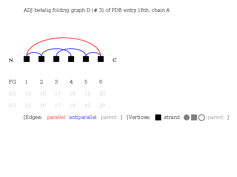
| 3 | D | 6 | EEEEEE | 17 | (e,2ae,1ae,-5pe,1ae,2ae,-2ae) | Fold D | Go to linnots |
Go to protein graph
Folding graph images
The images below show the folding graphs (connected components) of the protein graph. The folding graphs and their linear notations are used to power the motif search and other features of this database server. Note that folding graphs of size 1 (isolated vertices in the graph) are not listed here -- they would not be of any use. In the images, the following abbreviations are used: PG = protein graph, FG = folding graph, SQ = sequential in chain.
Fold number 0 (fold name: A)
The adj betalig folding graph A (#0) of PDB 1fnh chain A:
Number of first SSE of fold in parent graph: 4
Download the visualization of fold A in formats: [PNG] [SVG] [PDF]
Download the graph file of fold A in formats: [GML] [JSON] [XML (XGMML)]
Fold number 1 (fold name: B)
The adj betalig folding graph B (#1) of PDB 1fnh chain A:
Number of first SSE of fold in parent graph: 8
Download the visualization of fold B in formats: [PNG] [SVG] [PDF]
Download the graph file of fold B in formats: [GML] [JSON] [XML (XGMML)]
Fold number 2 (fold name: C)
The adj betalig folding graph C (#2) of PDB 1fnh chain A:
Number of first SSE of fold in parent graph: 11
Download the visualization of fold C in formats: [PNG] [SVG] [PDF]
Download the graph file of fold C in formats: [GML] [JSON] [XML (XGMML)]
Fold number 3 (fold name: D)
The adj betalig folding graph D (#3) of PDB 1fnh chain A:
Number of first SSE of fold in parent graph: 17
Download the visualization of fold D in formats: [PNG] [SVG] [PDF]
Download the graph file of fold D in formats: [GML] [JSON] [XML (XGMML)]